LCMS Separation of Impurities and Degradants
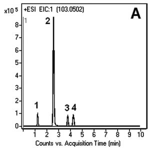
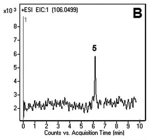
While two major impurities observed in this Cycloserine method were not directly identified, several possibilities can be suggested based on their m/z values in the Mass Spectrum and their relationship to the Serine structure. Serine was also identified but in very low abundance and only after the samples were several weeks old.
This study did not involve the development of a fully validated method; however a linear relationship following the equation 2E+6x – 280000 was obtained for the determination of Cycloserine over the concentration range of 0.2–1.0µg / mL having an R2 value of 0.993. The limit of detection is estimated to be 0.1µg / mL. The repeatability of the method, both inter and intra-day, is good.


Peak:
1. Unknown at m/z 285
2. Unknown at m/z 245
3. Cycloserine dimer at m/z 205.0931
4. Cycloserine at m/z 103.0502 (Fig. A)
5. Serine at m/z 106.0499 (Fig. B)
Method Conditions
Column: Cogent Diamond Hydride™, 4μm, 100Å
Catalog No.: 70000-15P-2
Dimensions: 2.1 x 150mm
Mobile Phase:
—A: DI Water / 0.1% Formic Acid (v/v)
—B: Acetonitrile / 0.1% Formic Acid (v/v)
Gradient:
| Time (minutes) | %B |
| 0 | 70 |
| 2 | 20 |
| 6 | 20 |
| 7 | 70 |
Post Time: 2 minutes
Flow rate: 0.4 mL / minute
Detection: ESI – POS – Agilent 6210 MSD TOF Mass Spectrometer
Injection vol.: 1μL
Sample Preparation: Stock solutions of the analytes were made in DI Water in the range of 0.2–0.7 mg / mL. All samples were filtered through a disposable 0.45µm Syringe Filter (MicroSolv Tech Corp.). Samples for injection were diluted 1:10 with 50:50 Solvent A:B mixture.
t0: 0.9 minutes
Note: Until recently, Cycloserine has not been in wide-spread use for the treatment of tuberculosis due to its toxicity. With more drug-resistant strains of TB emerging, Cycloserine treatment is becoming more common.
Attachment
No 168 Cycloserine pdf 0.3 Mb Download File


